


In a groundbreaking paper published in the prestigious journal Science, researchers from the University of Chicago have unveiled a revolutionary understanding of dinosaur preservation, detailing how the bodies of the duck-billed dinosaur Edmontosaurus annectens were transformed into exceptionally preserved "mummies" approximately 66 million years ago. This extraordinary preservation, termed "clay templating" by the scientific team, has yielded incredibly fine details of skin, scales, and even hooves, offering an unparalleled glimpse into the life of these Late Cretaceous creatures.
The process of clay templating, as described by the UChicago researchers, involved the rapid burial of the dinosaur carcasses. Following death and exposure, the outer soft tissues were preserved by a delicate clay coating that settled over the skeleton. This clay layer, remarkably thin—less than one-hundredth of an inch thick—acted as a precise mold, or template, capturing the intricate textures of the dinosaur’s exterior.
A Fleshed-Out Vision of a Lost World
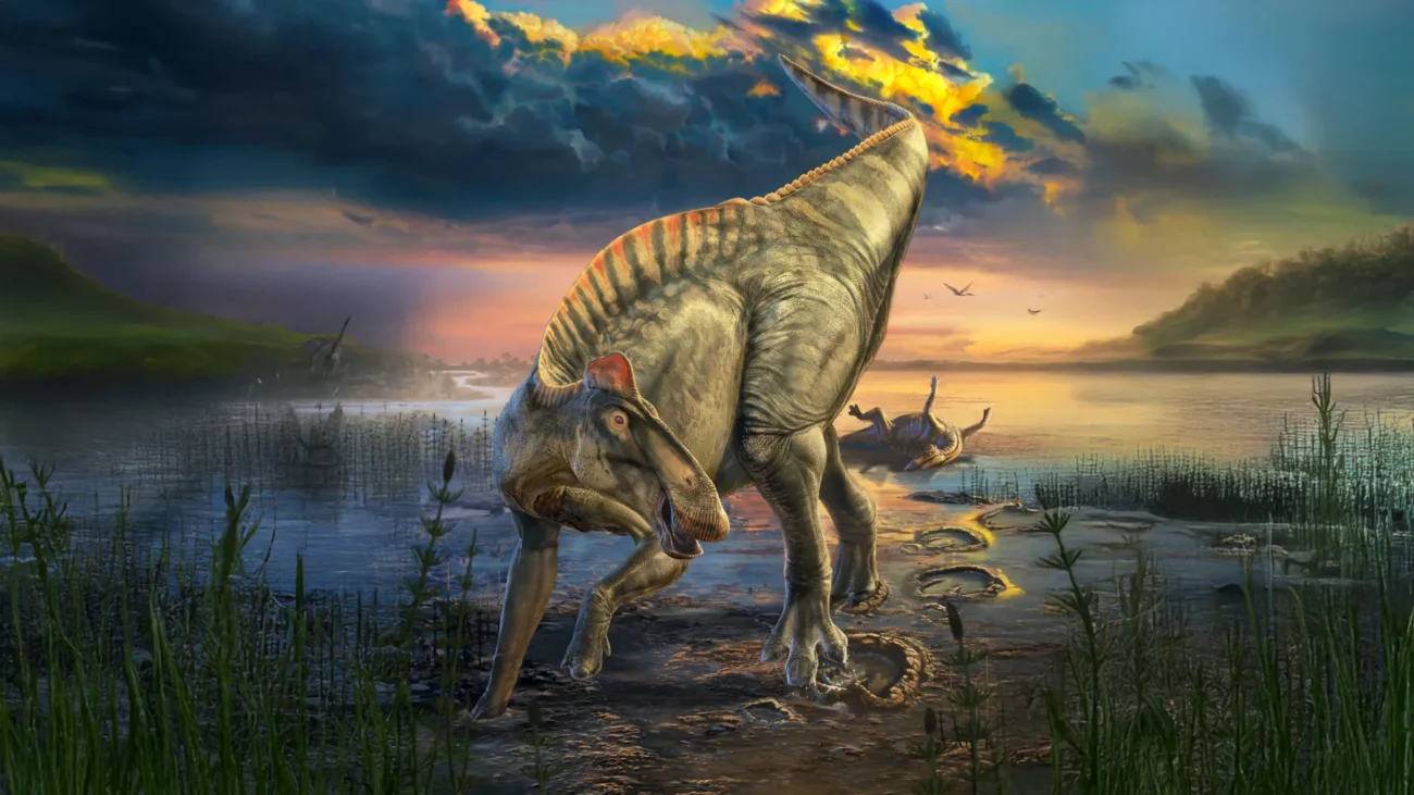
Leveraging advanced imaging techniques, including hospital-grade and micro-CT scans, the scientists have been able to reconstruct a remarkably detailed picture of the Edmontosaurus annectens in its living state. The digital reconstructions reveal a striking silhouette: a prominent crest running along the neck and torso, a distinctive row of spikes adorning the tail, and unique hooves that encased the toes. When these anatomical findings are correlated with fossilized footprints discovered in the same geological strata, the overall appearance and locomotion of this iconic duck-billed dinosaur, long a subject of scientific speculation but never before documented with such precision, become strikingly clear.
"It’s the first time we’ve had a complete, fleshed-out view of a large dinosaur that we can really feel confident about," stated senior author Paul Sereno, PhD, a Professor of Organismal Biology and Anatomy at the University of Chicago. He elaborated on the significance of the discovery, noting that the badlands of Wyoming, where the specimens were found, represent a unique "mummy zone" that continues to yield surprising fossil evidence from expeditions conducted over many years by university undergraduate teams.
Rediscovering Wyoming’s Dinosaur "Mummy Zone"
The scientific endeavor began with meticulous detective work, utilizing historical field photographs and geological records to retrace the exact locations in east-central Wyoming where several seminal dinosaur mummies were first unearthed in the early 1900s. Sereno and his team successfully mapped a concentrated "mummy zone" within these ancient riverbed deposits, which are characterized by stacked layers of river sands.
Within this geologically rich area, the researchers excavated two new Edmontosaurus mummies. One specimen represented a younger individual, while the other was somewhat older. Both of these specimens exhibit large, continuous patches of preserved external skin surface, providing crucial anatomical data necessary for constructing a comprehensive, lifelike profile of the animal.
Professor Sereno was keen to emphasize a key distinction between these dinosaur mummies and the human-created mummies found in ancient Egyptian tombs. He clarified that in the case of these fossils, none of the original organic material—such as skin or muscle tissue—remains. Instead, the preserved skin, spikes, and hooves are not biological remnants but rather an exceptionally thin clay coating that formed on the exterior of the carcass shortly after its burial. This coating, he explained, acted as a "mask, a template, a clay layer so thin you could blow it away." The preservation, he added, was the result of a "fluke event of preservation" where the clay was attracted to the outside of the carcass.
Ultra-Thin Clay Films: Capturing Dinosaur Skin in Three Dimensions
To fully understand the formation of these extraordinary fossils, the research team employed a comprehensive array of advanced imaging and analytical techniques. This included hospital-grade and micro-CT scans, which provide detailed internal and external structural information; thin sectioning, allowing for microscopic examination of the fossil material; X-ray spectroscopy, used to determine elemental composition; and detailed clay mineral analyses. A close study of the geological context, specifically the rock layers where the fossils were discovered, also played a crucial role. Collectively, these investigations pointed towards a specific sequence of geological and biological events that led to this exceptionally rare form of preservation.
The researchers have proposed a compelling model for the formation of these mummies. They theorize that after the dinosaurs died, their bodies likely underwent a period of desiccation, or drying, on the surface before being rapidly buried by sudden flash floods. A microbial film, which typically forms on the surface of decaying organic matter, is believed to have played a pivotal role. This film attracted clay particles from the surrounding wet sediment through electrostatic forces. This attraction resulted in the formation of a wafer-thin clay template that meticulously replicated the animal’s outer contours in three dimensions. Over vast geological timescales, the original soft tissues would have decayed and dissolved, leaving behind the fossilized clay impression alongside the skeleton.
The Meticulous Process of Digital Reconstruction
The fragility of these paper-thin clay layers necessitated a painstaking and meticulous preparation process. Tyler Keillor, the Fossil Lab manager and a co-author on the study, led extensive efforts to carefully expose these delicate surfaces without causing any damage. This phase of the research demanded a high level of precision and expertise.
Complementing the physical preparation, another team, led by postdoctoral scholar Evan Saitta, utilized sophisticated 3D surface imaging and CT scans. These advanced imaging methods were combined with comparative analyses of fossilized footprints from the same geological period. By meticulously tracing the preserved soft anatomy, examining the sediments both within and surrounding the mummy, and successfully matching the dinosaur’s hooves to a specific footprint, the team was able to reconstruct the animal’s limbs with remarkable accuracy. Digital artists then collaborated closely with the paleontologists, translating the scientific data into lifelike reconstructions that vividly illustrate how this duck-billed dinosaur would have appeared and moved as it traversed the muddy landscapes of the Late Cretaceous, near the very end of the age of dinosaurs.
Professor Sereno expressed his belief in the value of assembling a dedicated, interdisciplinary "dream team" to produce scientific research that is both rigorous and accessible to the general public. "We’ve never been able to look at the appearance of a large prehistoric reptile like this," he remarked, adding with a touch of seasonal humor, "and just in time for Halloween."
Unveiling the Crest, Spikes, Scales, and Thin Skin
Working with the two newly described mummies, the researchers were able to synthesize a complete and detailed outline of the Edmontosaurus annectens. "The two specimens complemented each other beautifully," Sereno explained. "For the first time, we could see the whole profile rather than scattered patches."
One of the most significant anatomical revelations was the discovery of a continuous feature running along the central dorsal line of the animal. This structure began as a fleshy crest adorning the neck and torso and transitioned into a single row of prominent spikes along the tail. Each spike was precisely aligned above a vertebra, fitting snugly with its neighbors, suggesting a potentially defensive or display function.
The study also provided unprecedented detail on the dinosaur’s scale patterns. The largest polygonal scales were observed along the lower body and tail, while the majority of the animal’s body was covered in remarkably small, pebble-like scales, measuring only 1 to 4 millimeters across. These scales are surprisingly tiny for a dinosaur that could reach lengths exceeding 40 feet, indicating a more delicate integument than previously imagined. Furthermore, the presence of fine wrinkles preserved over the ribcage suggests that the skin of this particular duck-billed dinosaur was relatively thin.
Hooves and Heel Pads on a "Hoofed" Dinosaur
Perhaps the most astonishing discovery emerged from the hind feet of the larger mummy: this dinosaur possessed hooves. The tips of each of its three hind toes were encased in a distinct wedge-shaped hoof with a flattened underside, bearing a striking resemblance to the hooves of modern horses.
To confirm the precise morphology of these feet in life, the researchers integrated CT scan data of the mummified feet with high-resolution 3D images of the most well-preserved duckbill footprint from the same geological period. Through careful alignment of the skeletal and soft-tissue impressions, they produced a detailed reconstruction of the hind foot. Intriguingly, unlike the forefoot, which appears to have made contact with the ground primarily through its hooves, the hind feet also featured a fleshy heel pad situated behind the hooves, suggesting a more complex weight-bearing mechanism.
Professor Sereno highlighted the profound significance of these findings, stating, "There are so many amazing ‘firsts’ preserved in these duck-billed mummies—the earliest hooves documented in a land vertebrate, the first confirmed hooved reptile, and the first hooved four-legged animal with different forelimb and hindlimb posture." These discoveries challenge previous assumptions about dinosaur anatomy and locomotion.
A New Toolkit for Dinosaur Soft-Tissue Research
Beyond the spectacular new anatomical details it has unveiled, this research offers a robust and practical framework for future investigations into dinosaur soft tissue preservation. The authors have meticulously outlined novel preparation techniques, established a clear set of terminology for describing soft structures and various scale types, and detailed a comprehensive imaging pathway from raw fossil specimen to fully rendered, lifelike model. Crucially, they have provided a scientifically grounded explanation for how dinosaur mummies can form under natural geological conditions.
The paper’s contribution extends beyond a series of isolated findings. It proposes a generalized model for dinosaur mummification, based on the principle of clay templating, which can now be applied and tested on other fossil discoveries exhibiting similar preservation characteristics.
Looking ahead, the research team has identified several key areas for future work. These include focused expeditions to locate additional specimens with this unique type of preservation in the same Wyoming rock layers and in other potentially analogous geological regions. Further biomechanical studies will now benefit from the availability of accurate external body outlines, enabling more precise analyses of locomotion and function. Additionally, complementary geochemical and sedimentological analyses are planned to better understand the environmental conditions under which clay templating is most likely to occur.
Professor Sereno concluded by reflecting on the comprehensive nature of the study, stating, "This may be the single best paper I’ve released. From field to lab to 3D reconstructions along with a suite of useful terms defined, it’s a tour de force, and it tells a coherent story about how these remarkable fossils come to be and what we can learn from them." This seminal work not only rewrites our understanding of Edmontosaurus but also provides a foundational methodology for unlocking further secrets of dinosaur soft tissue preservation, promising to reshape paleontological research for years to come.