


In a groundbreaking discovery published in the prestigious journal Science, researchers from the University of Chicago have unveiled a revolutionary process, termed "clay templating," that has preserved the intricate details of duck-billed dinosaurs, Edmontosaurus annectens, in astonishing "mummy-like" fossils. This remarkable preservation, dating back approximately 66 million years, has allowed scientists to reconstruct the appearance and anatomy of these ancient creatures with unprecedented accuracy, shedding new light on their morphology and locomotion.
The Genesis of Dinosaur Mummies: Clay Templating Unveiled
The key to this extraordinary fossil preservation lies in a rare geological phenomenon that the University of Chicago team has named "clay templating." This process, occurring in specific ancient riverine environments, involved the rapid burial of dinosaur carcasses, leading to the formation of an ultra-thin clay coating that acted as a precise mold of the animal’s outer tissues. This delicate clay mask, less than one-hundredth of an inch thick, captured the minute textures of skin, scales, and even hooves, providing a three-dimensional imprint after the original organic material had long since decomposed.
The research team, led by Professor Paul Sereno, a renowned paleontologist at the University of Chicago, utilized advanced imaging techniques to meticulously analyze these fossilized clay impressions. By combining high-resolution CT scans, micro-CT scans, and other sophisticated analytical methods, they were able to digitally reconstruct the three-dimensional form of the Edmontosaurus annectens. This allowed them to visualize features that had previously been a matter of speculation, such as a prominent crest along the neck and torso, a distinctive row of spikes adorning the tail, and the surprising presence of hooves enclosing the toes.
"It’s the first time we’ve had a complete, fleshed-out view of a large dinosaur that we can really feel confident about," stated Dr. Sereno, the senior author of the study. His enthusiasm underscores the significance of this discovery, which offers a tangible connection to the distant past. The badlands of Wyoming, a region historically rich in fossil finds, have proven to be a unique "mummy zone," a testament to the exceptional preservation conditions that have yielded surprises for decades.
Rediscovering Wyoming’s Prehistoric Treasure Trove: The "Mummy Zone"
The journey to understanding clay templating began with meticulous detective work. Dr. Sereno and his colleagues, armed with historical field photographs and an intimate knowledge of paleontological exploration, retraced the footsteps of early 20th-century paleontologists in east-central Wyoming. Their investigations led them to a concentrated area within the fossil-bearing rock layers—stacked river sands—that they identified as a "mummy zone." This zone, characterized by its exceptional preservation of soft tissues, has been a source of fascination and scientific inquiry for over a century.
Within this identified area, the team undertook new excavations, unearthing two remarkably well-preserved Edmontosaurus annectens specimens. One was a juvenile, and the other a more mature individual, both exhibiting large, continuous sections of their external skin. These fossils were crucial in providing the complete anatomical data needed to construct a comprehensive picture of the dinosaur’s form and appearance.
Dr. Sereno clarified a common misconception about these dinosaur "mummies," distinguishing them from the human-created mummies of ancient Egypt. "None of the original organic material is still present," he emphasized. Instead, the preserved skin, spikes, and hooves are not remnants of original tissue but rather an extremely thin clay coating that formed on the exterior of the carcass shortly after its entombment. This "mask," as he describes it, was an accidental yet perfect replica.
The Science Behind the Marvel: Ultra-Thin Clay Films in 3D
The formation of these extraordinary fossils has been elucidated through a rigorous scientific investigation employing a battery of advanced imaging and analytical techniques. The research team utilized hospital-grade CT scanners and micro-CT scanners to peer into the intricate structures of the fossils. Complementary analyses included the examination of thin sections of the fossilized material, X-ray spectroscopy to determine elemental composition, and detailed clay mineral analyses to understand the geological context. Crucially, a thorough study of the rock layers where the fossils were discovered provided vital clues about the depositional environment and the sequence of events that led to such rare preservation.
The proposed model of formation involves a sequence of natural processes:
- Drying and Rapid Burial: Following death, the dinosaur’s body would have been exposed to the elements, undergoing desiccation. Subsequently, sudden and violent flash floods would have rapidly buried the carcass within the ancient riverbed sediments.
- Microbial Attraction of Clay: A microbial film, likely a biofilm formed on the surface of the decomposing carcass, played a pivotal role. This film possessed electrostatic properties that attracted positively charged clay particles from the surrounding wet sediment.
- Clay Templating: Through these electrostatic forces, a thin, uniform layer of clay adhered to the outer surface of the carcass, creating a delicate but precise mold. This "clay template" captured the three-dimensional contours of the animal’s skin, scales, and other external features.
- Decomposition and Fossilization: Over geological timescales, the soft tissues within the clay mold decayed and disappeared. The clay template, however, remained intact, preserving the external morphology. Simultaneously, the underlying skeleton underwent the process of fossilization, becoming mineralized over millions of years.
Meticulous Reconstruction: From Fossil Fragments to Lifelike Recreations
Unveiling the delicate structures preserved within these fossils demanded exceptional care and precision. Tyler Keillor, the Fossil Lab manager at the University of Chicago and a co-author of the study, spearheaded the painstaking preparation of the specimens. His team dedicated countless hours to meticulously cleaning and exposing the fragile clay surfaces without causing any damage. This delicate work was essential to accessing the crucial information contained within the thin clay films.
Following the physical preparation, another group, led by postdoctoral scholar Evan Saitta, employed advanced 3D surface imaging and CT scans to digitally map the preserved soft anatomy. This detailed digital information was then cross-referenced with a comprehensive database of fossilized footprints from the same geological period. By meticulously aligning the reconstructed dinosaur feet with the fossilized trackways, the researchers were able to confirm the morphology of the hooves and their interaction with the substrate.
This integrated approach allowed for the creation of highly accurate, lifelike digital reconstructions. Working in close collaboration with digital artists, the scientific team brought the Edmontosaurus annectens to life, illustrating how it would have appeared and moved as it navigated the muddy landscapes near the close of the Cretaceous period. The visual representations are not mere artistic interpretations but are grounded in robust scientific data, offering a scientifically validated glimpse into the past.
"I believe it’s worth taking the time to assemble a dream team in order to generate science that can be appreciated by the general public," Dr. Sereno remarked. This sentiment highlights the dual mission of the research: advancing scientific understanding and making these discoveries accessible and engaging for a broader audience, especially with events like Halloween providing a natural context for interest in prehistoric creatures.
Unveiling the Duckbill’s Secrets: Crest, Spikes, Scales, and Thin Skin
The analysis of the two newly unearthed Edmontosaurus annectens mummies provided a remarkably complete picture of the dinosaur’s external anatomy. The complementary nature of the two specimens, as described by Dr. Sereno, was instrumental. "The two specimens complemented each other beautifully," he stated. "For the first time, we could see the whole profile rather than scattered patches."
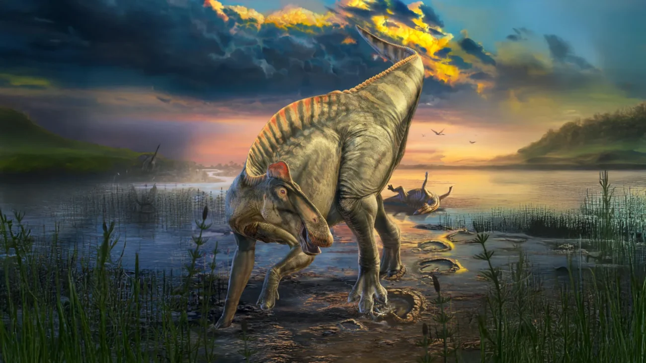
One of the most striking revelations was the discovery of a continuous feature running along the dorsal midline of the animal. This structure began as a fleshy crest that extended over the neck and torso. As it transitioned towards the posterior, it evolved into a single row of pointed spikes that ran along the tail. Each spike was precisely aligned with a vertebra, fitting snugly with its neighbors, suggesting a coordinated and potentially defensive or display-related function.
The research also provided significant insights into the dinosaur’s integumentary system, detailing its scale patterns. The largest, most prominent polygonal scales were found predominantly along the lower body and tail. In contrast, the majority of the animal’s body was covered in remarkably small, pebble-like scales, measuring only 1 to 4 millimeters in diameter. This fine scale covering is particularly surprising for a dinosaur that could reach lengths exceeding 40 feet, suggesting a smooth, streamlined appearance over much of its body. Furthermore, the presence of fine wrinkles preserved on the ribcage indicates that the skin of this duck-billed dinosaur was relatively thin, perhaps contributing to thermoregulation or flexibility.
A Hoofed Dinosaur: Hooves and Heel Pads on the Hind Feet
Perhaps the most unexpected and groundbreaking discovery emerged from the examination of the larger mummy’s hind feet: the presence of hooves. The study revealed that the tips of each of the three main hind toes were encased in a distinct, wedge-shaped hoof with a flat underside, exhibiting a striking resemblance to the hooves of modern equids, such as horses. This finding marks the earliest documented instance of hooves in a terrestrial vertebrate.
To further elucidate the function and appearance of these unique feet, the researchers combined high-resolution CT scans of the mummified hooves with detailed 3D images of the most well-preserved Edmontosaurus annectens footprint discovered from the same geological period. By carefully aligning the skeletal structure with the soft-tissue impressions from the footprint, they were able to generate a highly detailed reconstruction of the hind foot’s anatomy in life.
This reconstruction revealed a nuanced locomotor apparatus. While the forefeet made contact with the ground primarily through their hooves, the hind feet possessed a more complex structure. In addition to the hooves, the hind feet also featured a fleshy heel pad located behind the hooves, suggesting a specialized gait and weight distribution.
"There are so many amazing ‘firsts’ preserved in these duck-billed mummies," Dr. Sereno exclaimed. The study has provided evidence for the earliest hooves ever documented in a land vertebrate, confirmed the existence of a hooved reptile (as dinosaurs are reptiles), and identified the first known four-legged animal with distinct forelimb and hindlimb postures that include hooves. These discoveries fundamentally alter our understanding of dinosaur biomechanics and evolutionary adaptations.
A New Paradigm for Dinosaur Soft Tissue Research
Beyond the anatomical revelations, this research has established a robust and practical framework for future investigations into dinosaur soft tissues. The authors have meticulously outlined novel preparation techniques that allow for the delicate exposure of fragile fossilized soft tissues. They have also proposed a clear set of standardized terminology for describing soft structures and various scale types, which will facilitate consistent communication within the scientific community.
The paper presents a comprehensive, step-by-step imaging pathway, guiding researchers from the initial fossil specimen through advanced scanning and digital reconstruction to produce lifelike models. Furthermore, it offers a detailed "recipe" for how dinosaur mummies can form under natural conditions, providing a scientific model for the clay templating process. This generalized model can now be applied and tested on other fossil discoveries exhibiting similar preservation characteristics.
The implications of this work extend far beyond a single species. The methodology and the understanding of clay templating offer a new toolkit for paleontologists worldwide. This opens up the possibility of re-examining previously discovered specimens that may have been overlooked due to the subtlety of their soft tissue preservation.
The research team has outlined clear future research directions, including targeted searches for additional specimens exhibiting this exceptional preservation in the same Wyoming rock layers and in other geologically similar regions globally. The availability of accurate external body outlines will also enable more precise biomechanical studies, allowing scientists to model locomotion and physiology with greater fidelity. Additionally, complementary analyses will focus on understanding the specific environmental conditions and geological factors that favor the occurrence of clay templating, thereby refining the predictive power of the model.
"This may be the single best paper I’ve released," Dr. Sereno concluded, expressing profound satisfaction with the project’s comprehensive nature. "From field to lab to 3D reconstructions along with a suite of useful terms defined, it’s a tour de force, and it tells a coherent story about how these remarkable fossils come to be and what we can learn from them." This sentiment encapsulates the scientific rigor, collaborative spirit, and significant impact of this landmark study, which promises to redefine our understanding of dinosaur biology for years to come.
