


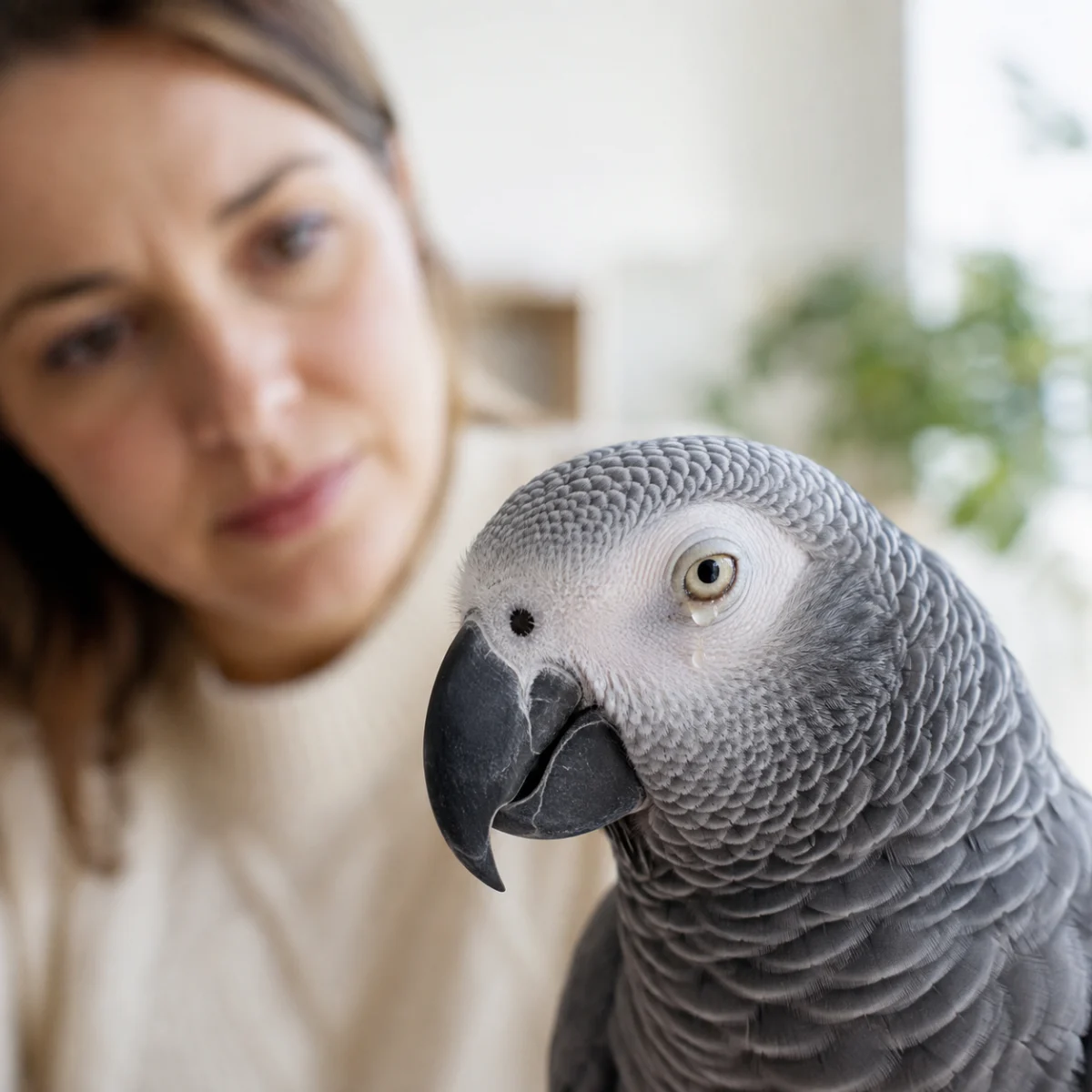
Recognizing subtle changes in a bird’s physical health is a critical responsibility for any avian companion owner. Among the most frequent concerns reported to veterinary clinics is the appearance of moisture, crusting, or discharge around the eyes. While some ocular moisture can be a transient, benign event related to daily grooming, persistent or abnormal discharge often serves as a primary indicator of underlying medical distress. Given the high metabolic rate and specialized anatomy of avian species, conditions that appear localized to the eye can often mask systemic infections, respiratory ailments, or nutritional deficiencies that require immediate professional intervention.
The Anatomy of Avian Ocular Health
To understand why eye discharge occurs, one must first consider the bird’s unique physiology. Unlike mammals, a bird’s eyes, nostrils, and sinuses are anatomically interconnected through a complex system of ducts and chambers. This proximity means that an issue manifesting as a "watery eye" is frequently a downstream effect of inflammation in the upper respiratory tract.
In a healthy state, a bird’s eyes should remain bright, alert, and clear. A brief period of moisture may occur following normal activities such as preening, bathing, or exposure to environmental particulates like household dust or feather dander. In these instances, the eye typically clears within minutes. However, when moisture persists, or when the discharge shifts from clear to opaque, it suggests that the body’s homeostatic mechanisms are being challenged by an external or internal irritant.
Chronology of Symptoms and Diagnostic Clues
The progression of eye-related illness generally follows a predictable timeline. In the initial phase, owners may notice occasional blinking or a slight sheen of moisture. If the cause is a minor foreign body—such as a piece of millet hull or a fiber from a toy—the bird’s natural tear production may flush the object out, returning the eye to a normal state.
If the discharge continues beyond this brief window, it enters a second phase of clinical significance. This is often characterized by the appearance of sticky or crusty buildup on the feathers surrounding the eyelid. At this stage, the bird may begin to exhibit behavioral changes: rubbing the side of its face against a perch or cage bar, squinting, or keeping the eye partially closed. These behaviors are clear indicators of ocular discomfort or pain.
The third, more critical phase involves the development of colored discharge—typically white, yellow, or green. This is a definitive sign of inflammation or bacterial infection. When accompanied by systemic symptoms such as fluffed feathers, lethargy, decreased appetite, or respiratory changes (e.g., clicking sounds or tail bobbing), the situation moves from a localized concern to an urgent medical priority.
Supporting Data and Clinical Analysis
Avian practitioners emphasize that the color and consistency of discharge act as clinical clues rather than diagnostic Certainties. Clinical studies have shown that the ocular surface is highly sensitive to environmental contaminants. According to data provided in Clinical Avian Medicine, environmental factors account for a significant percentage of primary ocular irritation. Specifically, birds housed in environments with high concentrations of aerosolized particles—such as scented candles, non-stick cookware fumes, or poor air quality—are statistically more likely to present with chronic, low-grade conjunctivitis.
Furthermore, nutritional status remains a cornerstone of ocular health. Research into the dietary requirements of companion parrots reveals that Vitamin A deficiency is a leading contributor to squamous metaplasia, a condition where the protective epithelial linings of the eyes and sinuses become compromised. This lack of structural integrity makes the ocular surface significantly more susceptible to opportunistic bacterial and fungal infections.
Professional Veterinary Protocols
When a bird is presented with ocular discharge, the veterinary objective is to differentiate between primary eye disease and secondary manifestations of systemic illness. The standard diagnostic pathway includes:
- Physical Examination: Assessing the symmetry of the face, the condition of the nares (nostrils), and the general demeanor of the patient.
- Ocular Staining: Utilizing fluorescent dyes to identify potential corneal ulcers or micro-scratches that are invisible to the naked eye.
- Cytology and Culture: Collecting samples of the discharge to determine the presence of specific pathogens, such as Chlamydia psittaci or various Gram-negative bacteria.
- Diagnostic Imaging: In cases where sinus involvement is suspected, radiographs or advanced imaging may be utilized to inspect the deeper bony structures of the skull.
The implication of these findings is that owners should strictly avoid the use of "home remedies" or over-the-counter human eye drops. Because the underlying causes—ranging from a physical scratch to a systemic bacterial infection—require diametrically opposed treatment protocols, using the wrong medication can mask symptoms while the infection advances, potentially leading to permanent vision loss or systemic sepsis.
The Role of Nutrition in Long-Term Prevention
While medical intervention is required for active infections, the role of preventative nutrition cannot be overstated. A balanced diet serves as the foundation for the immune system’s ability to protect the ocular tissues. Veterinarians often advise against exclusive seed-based diets, as they are notoriously low in the beta-carotene necessary for synthesizing Vitamin A.
Instead, a varied diet incorporating dark leafy greens, orange-fleshed vegetables, and high-quality formulated pellets provides the necessary biochemical support for the tissues lining the respiratory and ocular systems. It is important to note that while nutrition is a protective factor, it is not a cure for active injury or infection. Owners should view nutritional management as a long-term wellness strategy rather than an acute treatment plan.
Addressing Common Misconceptions
A common error among bird owners is the "watch and wait" approach. While it is reasonable to monitor a bird for a few minutes after a bath to see if moisture clears, extending this period to days can be detrimental. In the context of avian health, the "wait and see" strategy is often associated with delayed diagnosis of conditions that are highly treatable in their early stages but become complex and costly when allowed to progress.
Furthermore, owners often worry about the risk of infection spreading to other birds in the flock. While many causes of eye discharge are non-contagious, some, such as psittacosis or certain viral pathogens, are highly transmissible. Therefore, any bird exhibiting ocular symptoms should be considered a candidate for temporary quarantine until a veterinary professional can determine the specific etiology.
Summary of Best Practices
For the conscientious bird owner, the following steps are recommended when ocular abnormalities are observed:
- Observation: Document the frequency, duration, and color of the discharge, as well as any accompanying behavioral changes.
- Gentle Hygiene: If debris is present on the feathers, use only warm water and clean, soft gauze to remove loose material. Do not apply pressure or attempt to pry the eye open.
- Professional Consultation: Contact an avian veterinarian as soon as the discharge becomes recurring or is accompanied by other physical signs of illness.
- Environment Check: Evaluate the bird’s living space for potential irritants, including air quality, humidity levels, and common household toxins.
Ultimately, the eyes are a window into the broader health of an avian patient. Changes in ocular appearance should be treated as a serious communication from the bird that its health status has shifted. By prioritizing early veterinary intervention and focusing on long-term preventative care—including species-appropriate nutrition and a clean environment—owners can effectively mitigate the risks associated with ocular disease and ensure the longevity and well-being of their avian companions. Early, informed action remains the most effective tool in the preservation of ocular health.

