


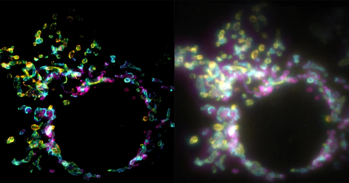
A groundbreaking advancement in optical imaging, known as spatial polarization-induced fluorescence fluctuation imaging (SPIFFI), has emerged as a transformative tool for cell biologists. Developed by a team of researchers at the Laboratory of Nanoscale Biology (LBEN) within the Swiss Federal Institute of Technology Lausanne (EPFL), this new technique bypasses the traditional limitations of super-resolution microscopy. By leveraging the intrinsic polarization of fluorescent molecules, SPIFFI allows for the capture of high-resolution images in a single exposure, effectively enabling the creation of detailed, high-speed videos of live cellular processes. This development marks a significant departure from conventional super-resolution methods, which historically required thousands of frames to reconstruct a single static image, rendering them largely unsuitable for studying the dynamic, ever-changing environment of a living cell.
The Evolution of Optical Microscopy and the Diffraction Limit
To understand the magnitude of the SPIFFI breakthrough, one must consider the fundamental constraints of optical microscopy. For over a century, light microscopy was governed by the Abbe diffraction limit, a physical constraint that prevents traditional optical systems from resolving details smaller than approximately half the wavelength of light—roughly 200 to 300 nanometers. In biological terms, this meant that many of the internal structures of a cell, such as the intricate architecture of the mitochondrial membrane or the movement of proteins along the cytoskeleton, remained blurred or entirely invisible.
The advent of super-resolution microscopy in the early 21st century—pioneered by researchers like Eric Betzig, Stefan Hell, and William Moerner, who were awarded the Nobel Prize in Chemistry in 2014—changed this landscape. Techniques such as STED (Stimulated Emission Depletion) and PALM/STORM (Photoactivated Localization Microscopy/Stochastic Optical Reconstruction Microscopy) allowed scientists to break the diffraction limit. However, these methods came with a significant cost: they were largely time-intensive. By relying on "temporal information"—essentially taking thousands of images of the same static sample to pinpoint the location of individual molecules—these techniques were fundamentally ill-suited for studying living cells. Because cells are in constant, rapid motion, a process that takes seconds or minutes to capture results in a "motion-blurred" image, effectively defeating the purpose of high-resolution imaging.
Understanding SPIFFI: The Mechanics of Polarization
The research published in the journal Nature by Wei Guo, Lely Feletti, and Aleksandra Radenovic introduces a paradigm shift. Instead of relying on time-sequenced snapshots, SPIFFI utilizes the physical properties of light emitted by fluorescent molecules. When a fluorophore (a fluorescent chemical compound) is excited by light, it does not emit photons in a purely random direction; rather, the emitted light is polarized. The orientation of this polarization is directly tied to the spatial orientation of the molecule itself.
The SPIFFI apparatus is designed to intercept this light and split it into four distinct polarization-sensitive channels. By comparing the variations in intensity across these channels simultaneously, the system can mathematically recover structural details that were previously lost to diffraction. Because the technique relies on the spatial distribution of light rather than the accumulation of thousands of temporal frames, the resolution enhancement happens in a single exposure.
"Essentially, previous approaches used temporal information to resolve spatial resolution, but this doesn’t work very well on living cells," explains Wei Guo, the first author of the study and a PhD student at LBEN. By shifting the burden from time to polarization, SPIFFI allows researchers to achieve a twofold increase in resolution—down to the 160-170 nanometer range—in every single frame.
Chronology and Development of the Technique
The development of SPIFFI was not an overnight success but the result of a concerted effort at EPFL to bridge the gap between high-speed imaging and high-resolution structural analysis. The research team focused on the following developmental milestones:
- Initial Conceptualization: The researchers identified that the untapped information contained in the polarization of fluorescent dyes could serve as a proxy for spatial density.
- Proof of Concept: The team designed an optical add-on module capable of splitting fluorescence into the four required polarization channels, ensuring that the hardware could be retrofitted onto existing commercial fluorescence microscopes.
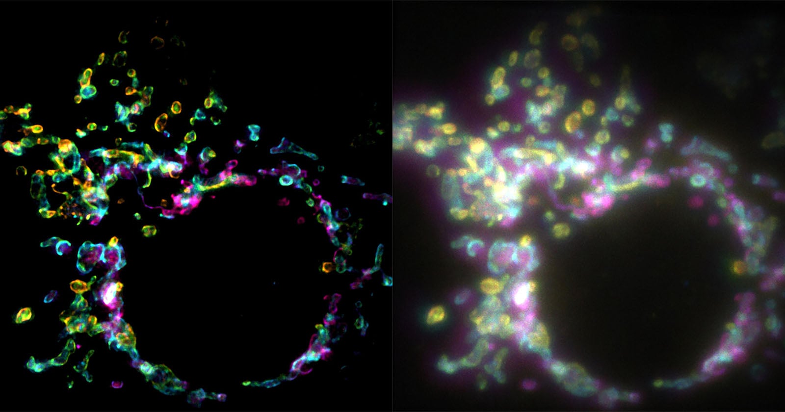
- Validation Phase: Using mitochondrial outer membranes as a model system, the team conducted side-by-side comparisons between traditional widefield microscopy and SPIFFI-enabled imaging. The results showed a drastic increase in contrast and resolution, confirming that the technique could resolve delicate structures that were previously rendered as indistinct blobs.
- Peer Review and Publication: Following rigorous testing to ensure the technique was robust across various biological samples, the findings were submitted to and published in Nature in September 2026.
Data-Driven Performance and Integration
One of the most compelling aspects of SPIFFI is its versatility. Beyond its standalone performance, which delivers roughly 160-170 nanometer resolution, the technique is designed to be compatible with existing post-processing algorithms. By integrating SPIFFI with traditional fluctuation-based super-resolution methods, the researchers successfully pushed the resolution boundary even further, achieving features as small as 80 nanometers.
This dual-layer approach is critical for the scientific community. It means that laboratories currently utilizing standard high-end fluorescence microscopes do not need to discard their equipment. Instead, they can integrate the SPIFFI optical hardware into their existing setups. This "modular" nature of the invention significantly lowers the barrier to entry for widespread adoption in academic and clinical research settings.
Furthermore, because every frame is "super-resolved," the technique enables the creation of true high-resolution time-lapse videos. This is a game-changer for cell biology, as it allows for the study of rapid, ephemeral events—such as cellular fusion, the transport of vesicles, or the remodeling of the cytoskeleton—in real-time. In previous experimental setups, such events would have been lost in the noise of the required multi-frame acquisition process.
Implications for Life Sciences and Medicine
The implications of SPIFFI for the broader scientific community are profound. By allowing researchers to peer into the living cell with unprecedented clarity and speed, the technique could unlock new insights into the pathogenesis of diseases. Many neurodegenerative conditions, for example, are characterized by the abnormal movement or misfolding of proteins within cells; being able to visualize these processes as they unfold could provide researchers with a new window into disease mechanisms.
Furthermore, the high-throughput capabilities of SPIFFI mean that it could be used in drug discovery and toxicology screening. Being able to quickly image a large number of cells at high resolution allows for faster statistical analysis of how specific compounds affect cellular structure over time.
"SPIFFI can capture fast-moving processes within cells, while enabling high-throughput, multi-dimensional imaging beyond the limits of conventional microscopes," notes Professor Aleksandra Radenovic. Her laboratory is already working on further refining the hardware, aiming to make the SPIFFI module more compact and user-friendly for non-specialists.
Future Outlook and Technical Challenges
While the initial results are promising, the researchers acknowledge that there is still work to be done. The current iteration of the hardware, while effective, requires precise calibration to ensure the four polarization channels are perfectly aligned. Future iterations are expected to focus on automated calibration and further miniaturization of the optics to allow for integration into even more compact microscope designs.
There is also the potential for SPIFFI to be applied to a wider range of biological dyes. Currently, the technique is most effective with fluorophores that exhibit strong, stable polarization characteristics. As the scientific community continues to develop brighter and more photostable fluorescent probes, the utility of SPIFFI is likely to expand, potentially allowing for even longer observation windows of living cells.
As the technology transitions from the laboratory setting to commercial development, the scientific community will likely watch closely to see if SPIFFI becomes a standard fixture in the modern bio-imaging toolkit. By successfully navigating the trade-off between speed and resolution, the team at EPFL has provided a new, practical path forward for one of the most critical challenges in cell biology. The ability to watch the "machinery of life" in high definition, without freezing it in time, represents a significant step toward a deeper, more dynamic understanding of biological systems at the nanoscale.

