


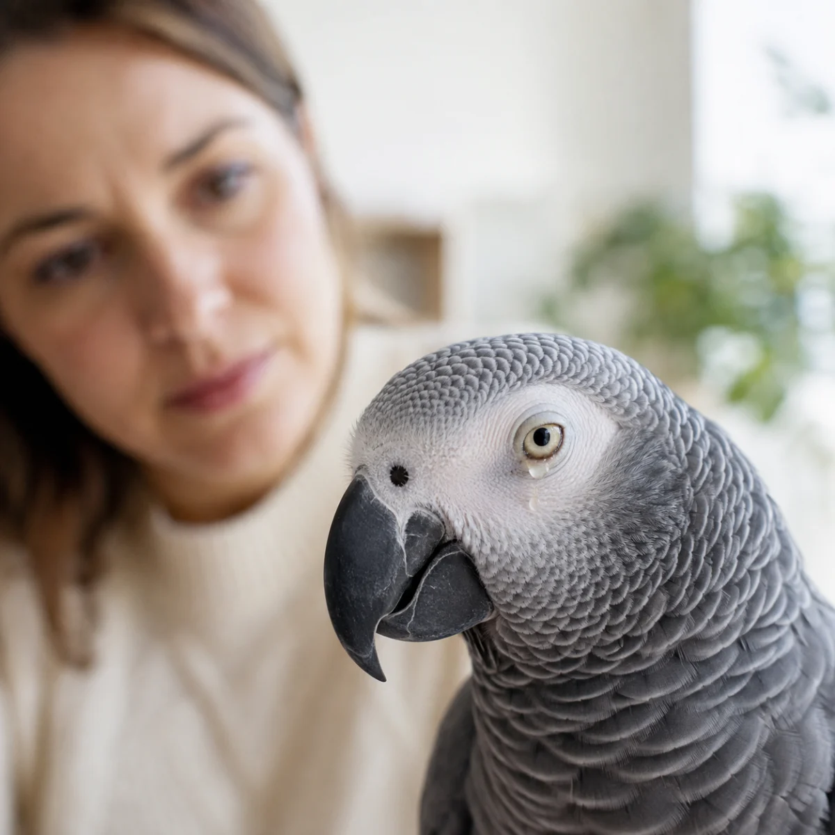
For many avian caregivers, the sight of a damp, crusted, or watery eye in a beloved parrot or songbird can trigger immediate alarm. While it is true that birds may occasionally exhibit minor moisture around the eyes due to grooming, dust, or environmental irritants, clinical experts emphasize that ocular discharge is a critical health indicator that should never be ignored if it persists. In the avian world, where birds often mask symptoms of illness until they are severe, a seemingly minor eye issue can be the first visible sign of a significant underlying condition, ranging from localized trauma to complex systemic infections.
The anatomy of a bird’s eye is exceptionally delicate and tightly integrated with the respiratory and sinus systems. Unlike mammals, where ocular issues are often isolated to the conjunctiva or cornea, an avian eye problem frequently serves as a window into the health of the bird’s entire cranial structure. Consequently, what may appear as a simple case of "morning tears" could, if left untreated, escalate into a life-threatening respiratory complication or permanent vision loss.
The Anatomy of Concern: Differentiating Normal from Pathological
To understand the urgency of eye discharge, one must first distinguish between transient moisture and true pathology. Brief, clear moisture following a bath or a vigorous preening session is typically a physiological response to physical activity or dust displacement. In healthy birds, such moisture clears within minutes, leaving the eye bright, clear, and fully open.
Conversely, discharge that lingers—specifically discharge that is cloudy, discolored (yellow or green), or accompanied by crusting on the surrounding feathers—is inherently pathological. Veterinary experts note that the presence of "morning tears" that fail to dissipate, or the appearance of sticky, mucoid buildup, suggests that the ocular surface is under stress. When a bird begins to exhibit secondary behaviors such as squinting, frequent blinking, or rubbing the affected side of its face against perches and cage bars, the situation has shifted from a minor irritation to a clinical concern requiring professional diagnostic intervention.
Chronology of Symptoms and Diagnostic Indicators
The progression of ocular distress in birds often follows a predictable, albeit rapid, timeline. In the initial phase, a bird may demonstrate mild discomfort characterized by intermittent blinking. If the underlying cause—such as a foreign body, a corneal scratch, or early-stage sinus inflammation—is not addressed, the condition enters a secondary phase within 24 to 48 hours. During this period, the discharge typically thickens, and the eyelid tissue may begin to swell, a phenomenon known as blepharoedema.
By the third phase of untreated symptoms, the bird’s systemic health may begin to decline. Because birds possess a high metabolic rate, infections can spread rapidly from the sinuses to the ocular cavity and beyond. It is at this stage that owners frequently observe the bird becoming "fluffed," lethargic, or showing a reduced interest in foraging. Data from avian clinical studies suggest that the earlier a veterinarian intervenes, the higher the success rate of non-invasive treatments. Waiting "a few days to see if it clears" is often the difference between a simple topical treatment and a multi-week regimen of oral antibiotics and intensive supportive care.
Supporting Data and Environmental Factors
Research into avian health consistently highlights that nutrition is a foundational pillar of eye health. Specifically, Vitamin A deficiency remains one of the most common nutritional contributors to ocular and respiratory morbidity in pet birds. Vitamin A is essential for the maintenance of epithelial tissues, which line the eyes, nostrils, and sinuses.
Birds on a strictly seed-based diet—which is notoriously low in Vitamin A—are statistically more prone to metaplasia, a condition where the tissues that normally protect the eye and sinuses become weakened and susceptible to bacterial colonization. While a change in diet will not "cure" an active infection, clinical observation proves that birds with a balanced, high-quality nutritional profile exhibit faster recovery times. Veterinarians strongly advise against the use of over-the-counter human eye drops or "home remedies," as these can inadvertently alter the pH of the bird’s eye or mask deeper, underlying structural damage that requires specialized ophthalmic equipment to visualize.
Clinical Perspectives and Veterinary Protocol
When an avian veterinarian performs an examination, the objective is to determine whether the discharge is primary (originating from the eye itself) or secondary (originating from the sinuses). This distinction is critical for determining the treatment path.
In a standard clinical assessment, the practitioner will:
- Ophthalmic Staining: Using fluorescein dye to identify microscopic corneal ulcers or scratches that are invisible to the naked eye.
- Sinus Evaluation: Checking for patency in the nares (nostrils) to determine if sinus congestion is pushing debris into the eye.
- Cytology/Culture: If the discharge is colored, a sample may be taken to identify specific bacterial or fungal pathogens.
- Imaging: In cases of chronic or recurrent discharge, radiographs may be required to assess the integrity of the infraorbital sinuses.
The consensus among veterinary professionals is that owners should never attempt to "force" the eye open or aggressively scrub away hardened discharge. Such actions can cause trauma to the sensitive nictitating membrane (the third eyelid) and may introduce further contaminants into the ocular sac. The safest course of action is to contact a qualified avian specialist immediately upon noting persistent symptoms.
Broader Impact: The "Silent" Nature of Avian Illness
The implications of ignoring eye discharge extend beyond the individual bird. Because many respiratory pathogens in birds, such as Chlamydia psittaci (psittacosis), can manifest with ocular symptoms, an untreated bird may pose a risk to other avian companions in the home. Furthermore, the psychosocial impact on a bird in pain is significant. Birds are prey species and naturally excel at hiding pain; by the time a bird allows an owner to notice that it is "squinting" or keeping one eye closed, it is often experiencing a level of discomfort that has already begun to compromise its appetite and immune response.
Actionable Steps for Bird Owners
For those currently observing discharge in their avian companions, the following framework is recommended by veterinary institutions:
- Observation: Document the timing, frequency, and color of the discharge. Note whether it is unilateral (one eye) or bilateral (both eyes).
- Documentation: Take clear, well-lit photos of the eye to share with your veterinary clinic. This allows the medical team to gauge the severity of the swelling before you even arrive.
- Hygiene: Maintain a pristine cage environment. Remove any loose debris or dust-heavy bedding that could exacerbate irritation.
- Immediate Consultation: If the bird appears weak, is holding the eye shut, or shows signs of respiratory distress (such as tail bobbing or audible clicking), treat this as an emergency.
In conclusion, while an occasional watery eye after a bath is rarely cause for alarm, persistent, recurring, or discolored discharge should be viewed as a clear signal that the bird’s internal equilibrium has been disrupted. The complexity of the avian facial structure—where the eyes and sinuses exist in an intimate, interconnected network—means that early intervention is the most effective tool in preventing chronic conditions. By maintaining a vigilant eye on your bird’s physical changes and prioritizing professional veterinary consultation over home-remedy speculation, you ensure the longest and healthiest life for your companion. The goal is to move beyond mere observation and into active, informed stewardship, ensuring that minor irritations do not become major medical emergencies.

